Discover more about the treatment of seven year old Christian, suffering from congenital flexural deformities of both distal interphalangeal joints (otherwise known as 'ballerina syndrome').
Christian Herbert, a seven year old gelding, arrived at the sanctuary as a foal in 2012 with congenital flexural deformities of both distal interphalangeal joints. Donkeys with this condition, often known as ballerina syndrome, are unable to fully weight bear through their solar surfaces and stand on their toes instead.
Although medical management and remedial farriery was initially commenced by the clinical team, the severity of the contraction in this donkey meant that this was not enough to resolve the deformities.
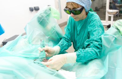
Christian subsequently underwent a deep digital flexor tenotomy at the mid metacarpal level of both forelimbs in a staged approach. The right forelimb was initially operated on followed by the left forelimb 7 days later.
These surgeries enabled Christian to weight bear normally on both feet and Christian was closely monitored throughout the subsequent years.
He remained sound and able to fully weight bear on both front feet until the age of 7, when the flexural deformity of his right distal interphalangeal joint recurred. This caused a recurrence of the abnormal weight distribution through the right front foot with only his toe being in contact with the ground.

Medical therapy and remedial farriery was subsequently commenced to promote lengthening of the musculotendinous unit of the right forelimb.
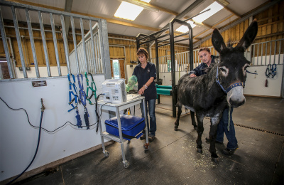
Our stable managers performed daily physiotherapy; gently encouraging extension of the distal aspect of the right forelimb and Christian was maintained on a consistent concrete surface during the day to further facilitate extension of the right forelimb.
The farriery interval was reduced to prevent excessive growth of the heel and remedial shoes utilising a silicone of hard consistency (Superfast, Vettec Hoof Care) were produced to elevate the toe region of the right front foot and further elongate the msuculotendinous unit.
Christian was maintained on an anti-inflammatory drug regime during this period to facilitate treatment.
Despite this treatment, Christian’s flexural deformity continued to deteriorate (Figure 2) and a decision to perform a further evaluation with a repeat tenotomy was made.

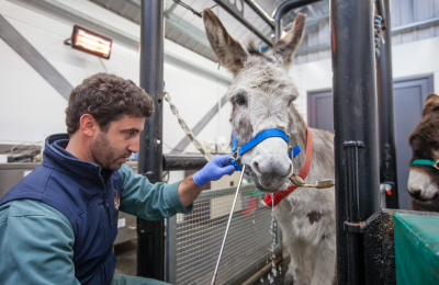
An ultrasonographic evaluation of the right metacarpal region was performed at our hospital which revealed extensive scar tissue and adhesions at the original surgical site.
A tenotomy of the deep digital flexor tendon of the right forelimb was repeated under general anaesthesia with the tenotomy being performed in a distal location to the original surgical site.
Christian showed complete resolution of his flexural deformity post-surgery and was discharged back to his farm.

Christian continued to make good progress and was maintained in a small hard area while remedial farriery was continued. A gradual increase in exercise intensity was performed and Christian eventually returned to his original group.